www.ptreview.co.uk
26
'21
Written on Modified on
BCN3D's 3D printing technology creates biomedical models that improve cancer surgeries
In Argentina, Dr Gustavo Nari has collaborated with engineering company Mirai3D to create biomedical models that significantly improve his surgical planning process thanks to the IDEX technology of his BCN3D Sigmax 3D printer.

BCN3D, a leading 3D printer manufacturer based in Barcelona (Spain), today announced that its 3D printing technology is helping surgeons to create biomedical models and apply them in their surgical procedures, particularly in cancer-related cases. A good example of this use of 3D printing technology is that of Dr Gustavo Nari in Argentina. He has collaborated with engineering company Mirai3D to develop and manufacture biomedical models that significantly improve his surgical planning process, using IDEX technology from his BCN3D Sigmax printer.
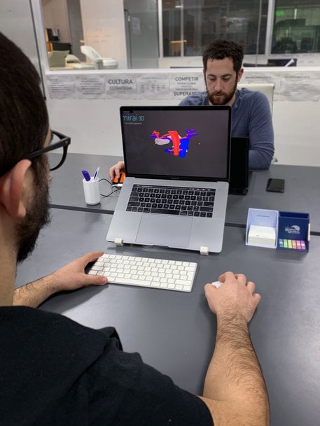
Dr. Gustavo Nari has collaborated with engineering startup Mirai3D to develop and fabricate biomedical models for an improved surgical planning process, making use of the IDEX technology in his BCN3D Sigmax printer.
All around the world, 3D printing is earning itself a reputation as a real asset to those in search of new ways to improve efficiency and precision in healthcare practices.
Across the pond, in the Argentinian city of Córdoba, Hospital Transito Caceres de Allende’s Dr. Gustavo Nari has teamed up with Mirai3D: a biomedical engineering startup that focuses its efforts on 3D printing, virtual reality, and advanced materials in the healthcare sector. Enlisting the help of the IDEX technology in a BCN3D Sigmax, they are creating biomodels to design surgical plans for a better anatomical understanding of clinical cases. Dr. Nari is experiencing first-hand the wonders this can bring to surgical procedures from start to finish.

So where does 3D printing come in?
Especially in cases of oncology, the surgeries that are required are intricate and complex, with doctors often having to navigate in and around vital organs. Preoperative surgical planning gives them a base from which to plan their routes of access and get a good idea of the step-by-step process. This also allows preemptive planning for any possible complications that may arise.
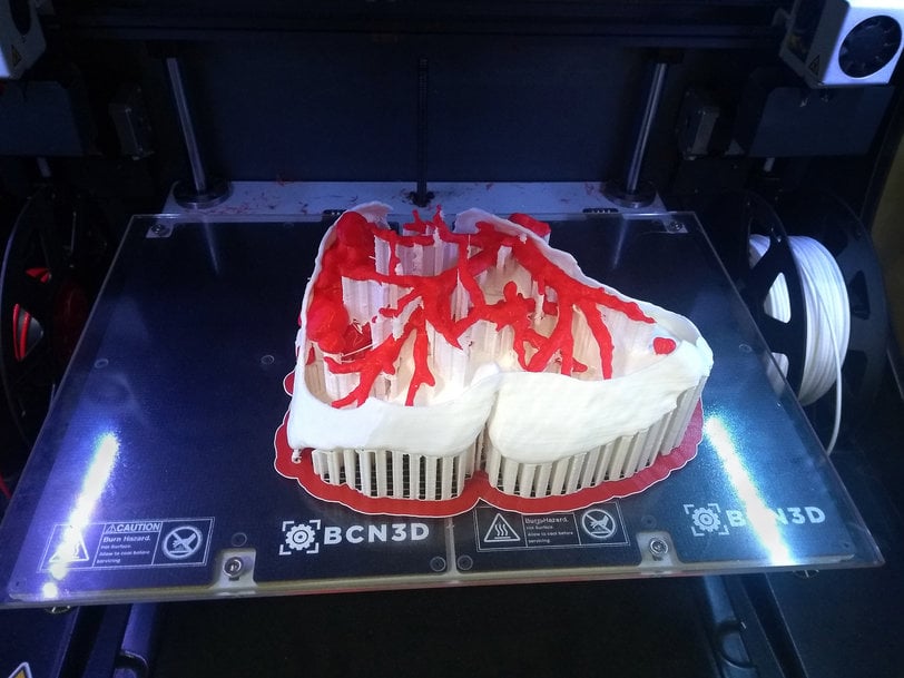
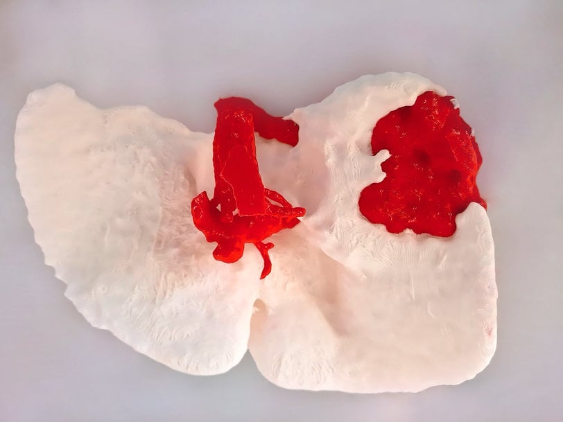
Combining X-rays and 3D printing technology allows doctors to use scans of the patient to produce a virtual model of the area of interest. This model can then be printed according to lifelike dimensions and sizings. IDEX technology is particularly advantageous here, as the model can be printed in different materials, to replicate different parts of the body such as bones or tissue. IDEX technology can also be used for a variety of colours, giving doctors a visual aid of the different areas. This means more training for doctors to build on their knowledge and have a thorough grasp on case-specific facts.
Furthermore, the models can be used to explain the procedure to the patient (or worried family members) in detail, and in an easy-to-follow way without the confusion of medical jargon, hopefully squashing any anxieties.
Patient with one liver metastasis
In this particular case, the patient is a 65-year-old woman, faced with one liver metastasis in the right lobe and another small one in the left. The patient’s complex clinical history meant that it was of the utmost importance to be meticulous and have rigorous control in this surgery. She had a history of double primary ovarian and colorectal tumor surgery and adjuvant treatment. Additionally, she had extremely low values of congenital thrombocytopenia (between 16 thousand and 25 thousand platelets when normal levels are above 150 thousand), meaning it was essential to reduce any large hemorrhages.
Using FFF 3D printing technology, PLA was printed at a resolution of 0.02mm. PLA was Dr. Nari’s material of choice, as it is cost-efficient and allows a variety of colours. Our Sigmax 3D printer allowed them to print two colours simultaneously, and led to a better visual representation and subsequent understanding. “I believe that in cases like these, 3D printing technology becomes an extremely helpful tool.” – Dr. Nari.

Biomodel printing
In the operating room, Dr. Nari proceeded to carry out a right hepatectomy together with a limited resection of the metastasis associated with the left lobe. At this point, the physical 3D model was used along with the virtual one for reference. The use of the biomodels meant that the doctor could view the problem of the tumor with its associated vascularization and physiological liver vascularization, facilitating a quicker surgery time. It provided an accurate reference of her vascular structure which was particularly important due to her pathology in relation to blood coagulation.
“Thanks to the use of this 3D model, the surgical time could be reduced by approximately 45 minutes, resulting in a total surgery time of 3 hours.”
The patient herself benefited from the experience with the 3D technology, as the assurance of the surgical team and their subsequent attention to detail warranted her safety. Furthermore, a shorter time span of surgery equates to a faster recovery, as the patient spends less time under the knife, which reduces the risk of complications or infections. “Thanks to the model, we entered the surgery with much more confidence. The knowledge of the vascular anatomy and its relationship with the tumour facilitates the surgical plan.”
There are plenty of benefits from incorporating additive manufacturing waiting to be discovered in the surgery room, as this clinical case and Dr. Nari’s results show. Dr. Nari is continuing his successful work with his Sigmax and reaping the benefits of cost and time efficiency, quick recovery times, and an increase in confidence in his team. As for Mirai3D, they are developing software for clinics and hospitals, while working towards their goal of opening an entire laboratory of 3D printing equipment. It’s hard to imagine a more beneficial way of using 3D printing technology!
www.bcn3d.com

