www.ptreview.co.uk
18
'25
Written on Modified on
Affordable LF-OCT System Achieves High-Speed Tissue Imaging
Off-the-shelf Mikrotron CoaXPress camera and BitFlow frame grabber enable 5000-fps, low-latency clinical OCT imaging with reduced cost and compact system design.
mikrotron.de
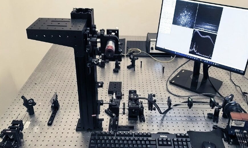
Set-up of the LF-OCT system (Photo courtesy of Oregon Health & Science University)
Optical Coherence Tomography (OCT) is a non-invasive imaging technology that uses light waves to produce high-resolution, cross-sectional images of biological tissues. OCT systems are most commonly used in eye care for early detection, diagnosis, and monitoring of various ocular conditions.
Line-field confocal optical coherence tomography (LF-OCT) combines principles from OCT and reflectance confocal microscopy. LF-OCT excels in scenarios requiring fine cellular resolution, such as early detection of skin cancers or treatment follow-up, where it can serve as a biopsy alternative. On the downside, LF-OCT systems are expensive, especially for individual practitioners or smaller clinics, as medical imaging equipment in this category often involves significant upfront costs.
Scientists at the Casey Eye Institute and the Department of Biomedical Engineering at Oregon Health & Science University (Portland, OR) recently devised a real-time LF-OCT system able to produce medically relevant images at 5000 frames per second using inexpensive SLED light sources and an off-the-shelf Mikrotron camera and BitFlow frame grabber. In doing so, the development team transformed a research-grade LF-OCT platform costing tens of thousands of dollars into an affordable, fully clinical-ready system. Through performance optimization and intelligent design modifications, engineers also achieved a significantly reduced footprint—delivering enhanced portability without compromising imaging capabilities. The streamlined configuration represents a critical advancement in bringing sophisticated OCT technology from the laboratory to point-of-care environments.
The clinical system leverages a precision-engineered hardware stack designed for maximum throughput and minimal latency. At its core, signal acquisition utilizes the Mikrotron EoSens 1.1CXP2 CMOS camera paired with a Bitflow Claxon CXP4 frame grabber, connected via a four-lane CoaXPress interface to achieve data transfer rates up to 12.5 Gbit/s—setting a new benchmark for clinical OCT systems.
The high acquisition speed makes capturing high-resolution en face images for different depths of focus much easier. Along with transmitting data, the highly deterministic, low-latency BitFlow Claxon frame grabber supplies 13W to the Mikrotron camera and provides a low-speed uplink, all over a single coaxial cable. The Mikrotron camera is equipped with a Zeiss Planar 1.4/85 M42-IR lens and configured in global shutter mode with resolution at 1024 x 500 pixels.
The system's computing platform is powered by Intel's Core i9-12900K processor, operating at peak frequencies of 5.20GHz and supported by 64 GB high-speed RAM. Parallel processing capabilities are amplified through the integration of an NVIDIA GeForce RTX 3090 graphics processing unit, enabling real-time execution of computationally intensive operations, including Fast Fourier Transform (FFT) calculations. A proprietary C/C++ processing framework orchestrates the complete imaging pipeline—from 5000 fps data acquisition through OCT processing, live image display, and secure data storage—with negligible processing delay. The optimized architecture delivers near-instantaneous visualization for clinical decision-making.
In testing, the Oregon Health & Science University system excelled in imaging cellular structures in both ex vivo and in vivo corneal tissues. The Mikrotron camera's ultra-high imaging speed reduced the need for sample stabilization and allowed the imaging of samples in rapid succession. At 5000 fps, many barriers associated with observing fast-changing processes were overcome. Another positive outcome was that imaging unstable samples or non-compliant individuals, such as very young children, became possible.
Although higher resolution and sensitivity may be required for specific analytical imaging goals, the system provides excellent support in a clinical setting, for instance, in monitoring anatomical structures during surgery.
The Oregon Health & Science University study aimed at reducing costs by using off-the-shelf components to develop an inexpensive LF-OCT for clinical applications. Less expensive components, including the integration of the Mikrotron camera and BitFlow frame grabber, plus the leveraging of the CoaXPress interface and parallel computing via the NVIDIA GPU, accelerate the potential commercialisation of the system to acquire, process, and display OCT data with low latency.
www.mikrotron.com